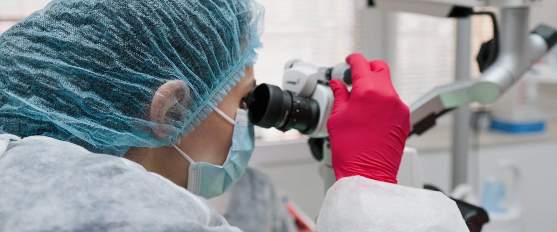
I have mentioned countless times that dentistry is constantly evolving. Given that this field involves working on such a small area—especially in endodontics, where we often rely more on our sense of touch than on actual visibility—there has been a constant search for methods to facilitate the work through better visibility. In other words, the need for magnification has become a priority. Initially, magnifying glasses were introduced, a magnification system worn by the dentist, with or without a light source. We will not deny the effectiveness of magnifying glasses, but what is truly revolutionary in the field of endodontics is a dental microscope.
The main advantages of using a dental microscope are that it provides a fixed, constant image position, is equipped with a light source whose intensity can be adjusted, and also has multiple magnification levels, thus allowing for highly precise procedures.
How a dental microscope works at each stage of endodontic treatment
- When it comes to accessing the pulp chamber (the cavity that houses the tooth's nerve), this is greatly facilitated by the use of a microscope. The increased visibility allows only the necessary amount of dental tissue to be removed in order to create the ideal geometry for access to the nerve, thus preserving the unaffected dental tissues that will provide additional strength to the tooth in the future.
- Repairing the opening holes of the root canals becomes much easier. There are many situations in which the entrance to the root canals is calcified or, in other words, blocked due to the mineralization of the dental pulp at this level. Among the possible causes of this phenomenon is the prolonged presence of untreated caries—the dental pulp practically retreats from the aggression represented by caries through mineralization. Without good visibility, it is almost impossible to permeate the dental canals, and in attempting to do so, unwanted accidents can occur that can compromise the success of the treatment.
- In addition, a dental microscope allows, through various magnification levels, visualization of the root canal trajectory and thus any remaining dental pulp in the case of treatment or filling material in the case of retreatment, facilitating their removal.
- Most importantly, a dental microscope allows visualization of foreign bodies in the root canals, such as broken needle fragments, and facilitates their removal.
- With the help of a microscope, possible cracks or fractures can be detected either coronally at the level of the walls or floor of the pulp chamber, or at the root level. In these practical situations, we can establish the prognosis of a tooth in terms of its retention in the arch.
- In the case of perforations at different levels or an apex (tip of the tooth), the increased visibility provided by the microscope allows them to be successfully closed with specific materials, increasing the chances of successful treatment.
- With the help of a microscope, a much more compact root canal filling can be achieved because there is control over the application of the material in the dental canals, which contributes significantly to the success of endodontic treatment.
The introduction of the dental microscope in dental practice, and more specifically in endodontics, has been truly revolutionary, and its effectiveness is undeniable. The advantages offered by increased visibility have contributed significantly to the increased success rate of endodontic treatments and thus to the longest possible preservation of teeth in the arch.